
Complex angioplasty
Coronary complex angioplasty has transformed the treatment of coronary artery disease. For many patients, a narrowed coronary artery can be treated safely with a balloon and coronary stent.
However, not every coronary blockage is straightforward.
Some patients have severely narrowed arteries with extensive calcium deposits. These are known as heavily calcified coronary lesions, and they can make angioplasty significantly more challenging.
In these situations, specialised techniques such as complex coronary angioplasty, rotational atherectomy (rotablation) and intravascular lithotripsy (IVL) may be used to prepare the artery before stent implantation.
What Is Coronary Complex Angioplasty?
Complex coronary angioplasty, also known as complex PCI (percutaneous coronary intervention), refers to angioplasty procedures that are technically more challenging than routine coronary stenting.
Complex PCI may be required when a patient has:
- Heavily calcified coronary arteries
- Chronic total occlusion (CTO)
- Long or multiple coronary blockages
- Complex bifurcation lesions
- Narrow or tortuous coronary arteries
- Previously failed angioplasty
- Multiple previous stents
- Diffuse coronary artery disease
- Left main or other high-risk coronary lesions
These procedures often require advanced equipment, specialised techniques and careful planning.
The goal is not simply to open the artery, but to create the best possible environment for the coronary stent to expand fully and remain effective over the long term.
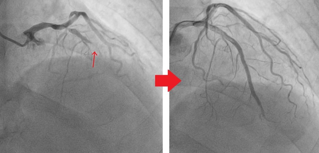
CTO mid LAD, crossed with Fielder-XTA then 2 stents at LAD
Why Is Calcium in the Coronary Artery a Problem?
Calcium can build up inside the walls of coronary arteries over many years.
Mild calcium may not cause major technical difficulties. However, severe calcification can make the artery extremely hard.
A conventional balloon may have difficulty crossing or expanding the lesion.
More importantly, even if a balloon and stent can be delivered, a severely calcified artery may prevent the stent from expanding properly.
Why is good stent expansion important?
A coronary stent is designed to act as a scaffold that keeps the artery open.
If the stent does not expand adequately, there may be a higher risk of complications, including:
- Stent under-expansion
- Blood clot formation inside the stent
- Re-narrowing of the artery
- Need for another procedure
Therefore, in heavily calcified lesions, calcium modification before stent implantation is often a crucial part of successful PCI.
What Is Rotablation?
Rotational atherectomy, commonly called rotablation, is an advanced technique used to modify heavily calcified coronary plaques.
During rotablation, a small diamond-coated burr is carefully advanced through the calcified coronary lesion.
The burr rotates at very high speed and modifies the hard calcium, allowing the lesion to become more compliant.
The objective is not usually to remove a large amount of plaque.
Instead, rotablation helps modify the calcium and facilitate balloon expansion and subsequent stent delivery.
When might rotablation be considered?
Rotablation can be particularly useful when:
- The lesion is severely calcified
- A balloon cannot cross the lesion
- A balloon cannot adequately expand
- The calcium is extensive or concentric
- Stent delivery is expected to be difficult
- Previous balloon-based calcium modification has been unsuccessful
Rotablation requires significant operator experience because the procedure involves specialised equipment and careful control of the burr, guidewire and coronary anatomy.
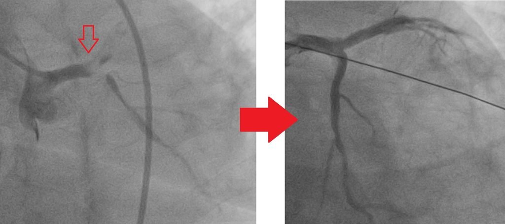
CTO LAD with Subtotal LCx, crossed with Conquest Pro 12, T stenting LM/LAD/LCx in high risk EF 25% with IABP
What Is Intravascular Lithotripsy (IVL)?
Another important technology for treating heavily calcified coronary arteries is intravascular lithotripsy (IVL).
IVL uses a specialised balloon catheter that delivers controlled acoustic pressure waves inside the coronary artery.
These pressure waves can create fractures within the calcium.
Once the calcium is modified, the artery becomes more capable of expanding with balloon inflation.
The final objective is to allow the coronary stent to achieve better expansion and apposition.
Rotablation vs IVL: What Is the Difference?
Both techniques are designed to modify calcium, but they work differently.
| Feature | Rotablation | IVL |
|---|---|---|
| Main mechanism | High-speed rotating diamond-coated burr | Acoustic pressure waves |
| Primary purpose | Modify severe calcium | Fracture/modulate calcium |
| Balloon required | No | Yes |
| Useful for undilatable lesions | Yes | Yes, if balloon can cross |
| Useful when lesion cannot be crossed | Often useful | May not be possible if IVL balloon cannot cross |
| Main strength | Excellent for severe, difficult calcium and balloon-uncrossable lesions | Controlled calcium modification with balloon-based therapy |
The choice between rotablation and IVL depends on the location, severity and distribution of calcium, lesion anatomy, balloon crossing and expansion, and the overall complexity of the PCI.
In selected cases, more than one calcium-modification strategy may be required.
How Do Cardiologists Know How Much Calcium Is Present?
Coronary angiography provides important information, but angiography alone may not fully define the extent of calcium.
Advanced intravascular imaging can provide much more detailed information.
Intravascular Ultrasound (IVUS)
IVUS uses ultrasound from inside the coronary artery to assess:
- Vessel size
- Plaque burden
- Calcium
- Stent expansion
- Stent apposition
Optical Coherence Tomography (OCT)
OCT uses near-infrared light to provide high-resolution images of the coronary artery.
It can help assess the characteristics and distribution of calcium and evaluate the final stent result.
In complex PCI, intravascular imaging can therefore help with planning, calcium modification and optimisation of stent implantation.

What Happens During Complex Angioplasty?
Every case is different, but a complex PCI involving severe calcification may follow several important steps.
1. Careful assessment
The coronary angiogram is reviewed to determine the severity and complexity of the blockage.
Where appropriate, IVUS or OCT may be used to understand the vessel and calcium more accurately.
2. Crossing the lesion
A specialised coronary guidewire is carefully advanced across the narrowed segment.
In complex lesions, this can sometimes be one of the most technically challenging parts of the procedure.
3. Calcium modification
Depending on the anatomy, the cardiologist may use:
- Rotablation
- IVL
- High-pressure or specialised balloons
- Other calcium-modification techniques
4. Balloon preparation
Once the calcium has been modified, balloon angioplasty is performed to prepare the artery for stent implantation.
5. Coronary stent implantation
A drug-eluting stent is positioned across the diseased segment and expanded.
6. Stent optimisation
The final result is carefully assessed.
IVUS or OCT may be used to confirm that the stent has expanded adequately and is properly positioned against the artery wall.
This final step is particularly important in complex PCI.
Is Rotablation or IVL Better?
There is no single technique that is best for every patient.
The choice depends on the specific coronary anatomy.
For example, rotablation may be particularly useful when the lesion is extremely hard or when a balloon cannot cross the blockage.
IVL can be particularly attractive when the balloon can cross the lesion but the calcium prevents adequate expansion.
In some complex cases, techniques may be combined.
The important principle is:
The treatment should be tailored to the anatomy rather than choosing one technology for every patient.

Is Complex Angioplasty Safe?
Modern complex PCI techniques have significantly expanded the range of coronary lesions that can be treated percutaneously.
However, complex angioplasty is still more technically demanding than routine PCI.
Potential complications include:
- Coronary artery dissection
- Coronary perforation
- Slow or no-reflow
- Vessel closure
- Bleeding or vascular complications
- Heart attack
- Abnormal heart rhythm
- Kidney injury related to contrast
- Emergency surgery in rare situations
The risk depends on the patient’s overall condition and the complexity of the coronary anatomy.
For this reason, complex PCI should be performed by an experienced interventional cardiology team with appropriate equipment and support.
Can Heavily Calcified Coronary Arteries Be Treated Without Bypass Surgery?
In selected patients, yes.
The availability of advanced PCI techniques means that some patients with complex coronary disease can be treated using minimally invasive catheter-based procedures.
However, PCI and coronary artery bypass surgery (CABG) are not competing treatments in every situation.
For patients with extensive multivessel disease, left main disease, diabetes or other high-risk anatomical features, CABG may provide better long-term outcomes in selected circumstances.
The decision should therefore be individualised after reviewing the patient’s symptoms, coronary anatomy, heart function, medical conditions and surgical risk.
Why Experience Matters in Complex PCI
Complex coronary angioplasty requires more than simply having advanced equipment.
Successful treatment depends on:
- Detailed assessment of coronary anatomy
- Appropriate guide catheter and guidewire selection
- Understanding of calcium distribution
- Choosing the correct calcium-modification strategy
- Careful balloon and stent sizing
- Intravascular imaging when appropriate
- Management of potential complications
- Appropriate patient selection
Rotablation and IVL are powerful technologies, but they are tools within a broader complex PCI strategy.
The ultimate goal is always the same: to restore blood flow safely while achieving an optimal and durable stent result.
Take-Home Message
Heavily calcified coronary arteries can make angioplasty significantly more challenging.
Fortunately, advances in interventional cardiology have provided several techniques to overcome these difficult lesions.
Rotablation uses a high-speed diamond-coated burr to modify severe calcium, while intravascular lithotripsy (IVL) uses acoustic pressure waves to fracture calcium within the artery.
When combined with modern balloons, coronary stents and intravascular imaging such as IVUS and OCT, these technologies can allow experienced interventional cardiologists to treat many patients with complex coronary artery disease.
If you have been told that your coronary arteries are “heavily calcified,” “too hard for a balloon,” or “difficult to stent,” it may be worthwhile to discuss whether advanced calcium-modification techniques such as rotablation or IVL are appropriate for your particular anatomy.
Complex coronary disease requires an individualised treatment strategy. The right technique depends on the patient’s coronary anatomy, clinical condition and procedural risk.
Do contact Dr Lim Wei Juan Consultant Cardiologist at KPJ Johor Specialist Hospital